
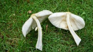
Green But Not Gold: The Deceptive Beauty of Chlorophyllum Molybdites
About This Article: Ready to meet the Shrek of the mushroom kingdom? Dive into our...
Keep Reading...
The Golden Princess: Breeding and Cultivating Agaricus Subrufescens
Welcome to the captivating world of mushroom cultivation, where nature’s treasures unravel in enchanting forms....
Keep Reading...
RECIPE: Pizza with Porcini Mushrooms, Sausage, and Truffle Sauce
Introduction: Unleash the flavors of the forest onto your dinner table with this Pizza featuring...
Keep Reading...
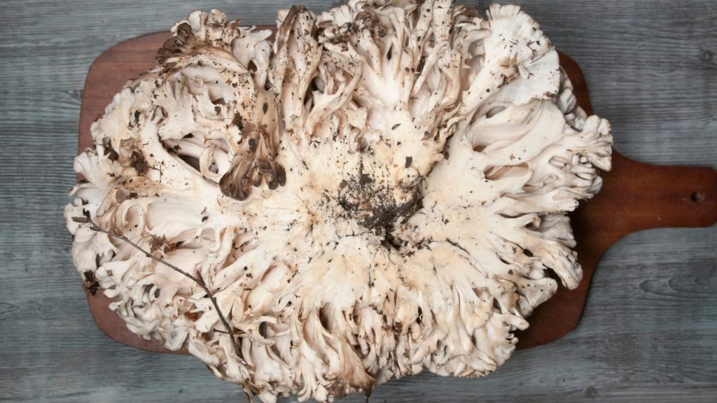
Cultivating Your Kingdom: A Guide to Growing King Oyster Mushrooms
The King Oyster Mushroom, also known as Pleurotus Eryngii, is the regal counterpart to the...
Keep Reading...